『物有光』,物之『表面』常為『光子 』『交互作用』的『時空』 ,『光線』魚貫各向進出的場域。所謂『成像法則』,『光子』之眾聚形成者也。因此『物點』對應『像點』,說明其『訊噪比』
訊號雜訊比
訊號雜訊比(英語:Signal-to-noise ratio,縮寫為SNR或S/N)是科學和工程中所用的一種度量,用於比較所需訊號的程度與背景雜訊的程度。其定義為訊號功率與雜訊功率的比率,以分貝(dB)為單位表示。大於比率1:1(高於0分貝)表示訊號多於雜訊。訊號雜訊比通常用於描述電子訊號,也可以應用在各種形式的訊號,比如冰芯內的同位素量,或細胞間的生物化學訊號。
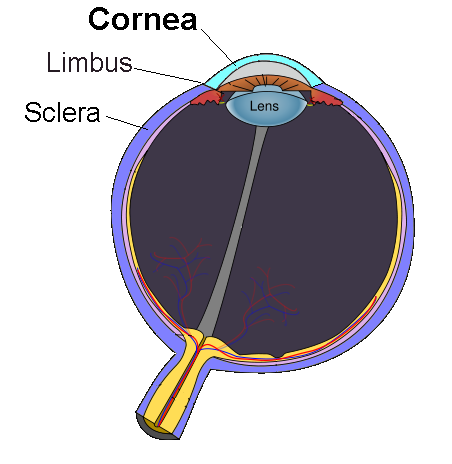
具『優勢』矣。這『光』  依理循徑,始於『眼角膜』
欲曉眼睛的光學原理,何不就沿著光徑
角膜→房水【前室】→虹膜【瞳孔】→晶狀體→玻璃體【後部】→視網膜
,從『角膜』之『屈光』能力開始。先讀維基百科詞條︰
角膜
角膜又稱黑睛,是眼睛最前面的透明部分,覆蓋虹膜、瞳孔及前房 ,並為眼睛提供大部分屈光力。加上晶狀體的屈光力,光線便可準確地聚焦在視網膜上構成影像。在人眼的折光系統中,角膜的折光能力是最強的,因為它直接和空氣接觸[1]。
角膜有十分敏感的神經末梢,如有外物接觸角膜,眼瞼便會不由自主地合上以保護眼睛。為了保持透明,角膜並沒有血管,透過淚液及房水獲取養份及氧氣。
人類角膜直徑約11.5毫米,中心厚度約有0.5至0.6毫米,邊緣厚度則約0.6至0.8毫米。
角膜過凸是真性近視的一個重要特徵,因此也是角膜塑形鏡與雷射手術(LASIK等)防治真性[近視]方法作用的部位[2]。
人工眼角膜
臺灣生技廠商在2005年開始投入上億經費研究,將臺灣鯛魚鱗片製成人工眼角膜,由於人和魚沒有共同疾病,不會有傳染病的疑慮,使用上安全性較高。
Cornea
The cornea is the transparent front part of the eye that covers the iris, pupil, and anterior chamber. The cornea, with the anterior chamber and lens, refracts light, with the cornea accounting for approximately two-thirds of the eye’s total optical power.[1][2] In humans, the refractive power of the cornea is approximately 43 dioptres.[3] While the cornea contributes most of the eye’s focusing power, its focus is fixed. The curvature of the lens, on the other hand, can be adjusted to “tune” the focus depending upon the object’s distance. Medical terms related to the cornea often start with the prefix “kerat-” from the Greek word κέρας, horn.
Structure
The cornea has unmyelinated nerve endings sensitive to touch, temperature and chemicals; a touch of the cornea causes an involuntary reflex to close the eyelid. Because transparency is of prime importance the cornea does not have blood vessels; it receives nutrients via diffusion from the tear fluid through the outside surface and the aqueous humour through the inside surface, and also from neurotrophins supplied by nerve fibres that innervate it. In humans, the cornea has a diameter of about 11.5 mm and a thickness of 0.5–0.6 mm in the center and 0.6–0.8 mm at the periphery. Transparency, avascularity, the presence of immature resident immune cells, and immunologic privilege makes the cornea a very special tissue. The cornea has no blood supply; it gets oxygen directly through the air. Oxygen first dissolves in the tears and then diffuses throughout the cornea to keep it healthy.[4]
It borders with the sclera by the corneal limbus.
The most abundant soluble protein in mammalian cornea is albumin.[5]
In lampreys, the cornea is solely an extension of the sclera, and is separate from the skin above it, but in more advanced vertebrates it is always fused with the skin to form a single structure, albeit one composed of multiple layers. In fish, and aquatic vertebrates in general, the cornea plays no role in focusing light, since it has virtually the same refractive index as water.[6]
─── 摘自《光的世界︰【□○閱讀】話眼睛《三》》
終至『視網膜』能二維成像  哩
哩
抵達視覺神經網絡之界面
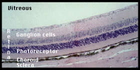
視網膜
視網膜又稱視衣,是脊椎動物和一些頭足綱動物眼球後部的一層非常薄的細胞層。它是眼睛裏面將光轉化為神經信號的部分。
視網膜含有可以感受光的視杆細胞和視錐細胞。這些細胞將它們感受到的光轉化為神經信號。這些信號被視網膜上的其它神經細胞處理後演化為視網膜神經節細胞的動作電位。視網膜神經節細胞的軸突組成視神經。視網膜不但有感光的作用,它在視覺中也有重要作用。在形態形成的過程中,視網膜和視神經是從腦中延伸出來的。
視網膜上的血管的結構每個人都不一樣,因此可以用來做生物特徵識別。
視網膜
解剖學
人的視網膜分10個層(由最外到最內):
- 視網膜色素上皮
- 感光層——包括視桿細胞及視錐細胞
- 外界膜——這個層隔開感光細胞的內部與其細胞核
- 外核層——又稱外顆粒層,由光感受器細胞核組成
- 外叢狀層——由光感受器細胞的軸突及雙極細胞(en:Bipolar neuron)樹突水平細胞突起組成,它們之間的接觸稱為突觸
- 內核層——又稱內顆粒層,由雙極細胞、水平細胞、無長突細胞、Muller細胞的胞核組成
- 內叢狀層——主要由雙極細胞的軸突及神經節細胞的樹突組成,並以突觸形式相接觸
- 神經節細胞層——這個層含有神經節細胞的細胞核,視神經從這裡開始
- 神經纖維層——主要為神經節細胞的軸突
- 內界膜
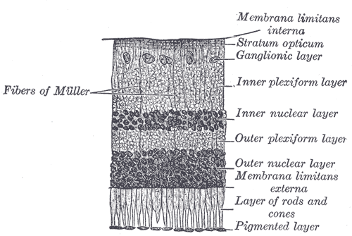
視網膜的不同層級
Retina
The retina (UK /ˈrɛtɪnə/ RET-i-nə, US /ˈrɛtᵊnə/ RET-(ə-)nə, pl. retinae, /ˈrɛtiniː/; from Latin rēte, meaning “net”) is the third and inner coat of the eye which is a light-sensitive layer of tissue. The optics of the eye create an image of the visual world on the retina (through the cornea and lens), which serves much the same function as the film in a camera. Light striking the retina initiates a cascade of chemical and electrical events that ultimately trigger nerve impulses. These are sent to various visual centres of the brain through the fibres of the optic nerve.
In vertebrate embryonic development, the retina and the optic nerve originate as outgrowths of the developing brain, specifically the embryonic diencephalon; thus, the retina is considered part of the central nervous system (CNS) and is actually brain tissue.[1][2] It is the only part of the CNS that can be visualized non-invasively.
The retina is a layered structure with several layers of neurons interconnected by synapses. The only neurons that are directly sensitive to light are the photoreceptor cells. For vision, these are of two types: the rods and cones. Rods function mainly in dim light and provide black-and-white vision while cones support the perception of colour. A third type of photoreceptor, the photosensitive ganglion cells, is important for entrainment and reflexive responses to the brightness of light.
Neural signals from the rods and cones undergo processing by other neurons of the retina. The output takes the form of action potentials in retinal ganglion cells whose axons form the optic nerve. Several important features of visual perception can be traced to the retinal encoding and processing of light.
─── 摘自《光的世界︰【□○閱讀】話眼睛《十五》》
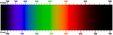
熟料『視生色』,不同『波長』  形成之『光譜』,竟有『感受』差異的
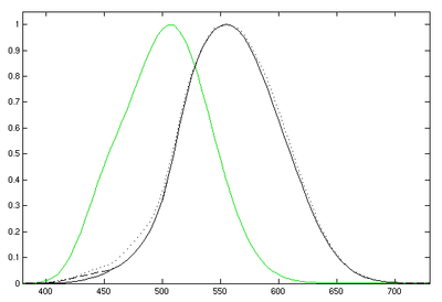
光度函數
光度函數或相對視見函數為人眼對不同波長光的平均視覺靈敏度,可用於將輻射能量轉化為可見光的計算。它並非在所有情況下都完全準確,而是一個以實驗方式得到的平均值。經由國際照明委員會(CIE)確認後,現已成為所有色彩科學使用的標準函數,並進一步成為CIE1931色彩空間的標準顏色匹配函數之一。
Luminosity function
The luminosity function or luminous efficiency function describes the average spectral sensitivity of human visual perception of brightness. It is based on subjective judgements of which of a pair of different-colored lights is brighter, to describe relative sensitivity to light of different wavelengths. It should not be considered perfectly accurate in every case, but it is a very good representation of visual sensitivity of the human eye and it is valuable as a baseline for experimental purposes.
The CIE luminosity function y(λ) or V(λ) is a standard function established by the Commission Internationale de l’Éclairage (CIE) and may be used to convert radiant energy into luminous (i.e., visible) energy. It also forms the central color matching function in the CIE 1931 color space.
於是乎有『彩色』的『大千世界』耶!!
Details
There are two luminosity functions in common use. For everyday light levels, the photopic luminosity function best approximates the response of the human eye. For low light levels, the response of the human eye changes, and the scotopic curve applies. The photopic curve is the CIE standard curve used in the CIE 1931 color space.
The luminous flux (or visible power) in a light source is defined by the photopic luminosity function. The following equation calculates the total luminous flux in a source of light:

where
- Φv is the luminous flux, in lumens;
- Φe,λ is the spectral radiant flux, in watts per nanometre;
- y(λ), also known as V(λ), is the luminosity function, dimensionless;
- λ is the wavelength, in nanometres.
Formally, the integral is the inner product of the luminosity function with the light spectrum.[1] In practice, the integral is replaced by a sum over discrete wavelengths for which tabulated values of the luminosity function are available. The CIE distributes standard tables with luminosity function values at 5 nm intervals from 380 nm to 780 nm.[cie 1]
The standard luminosity function is normalized to a peak value of unity at 555 nm (see luminous coefficient). The value of the constant in front of the integral is usually rounded off to 683 lm/W. The small excess fractional value comes from the slight mismatch between the definition of the lumen and the peak of the luminosity function. The lumen is defined to be unity for a radiant energy of 1/683 W at a frequency of 540 THz, which corresponds to a standard air wavelength of 555.016 nm rather than 555 nm, which is the peak of the luminosity curve. The value of y(λ) is 0.999,997 at 555.016 nm, so that a value of 683/0.999,997 = 683.002 is the multiplicative constant.[2]
The number 683 is connected to the modern (1979) definition of the candela, the unit of luminous intensity.[cie 2] This arbitrary number made the new definition give numbers equivalent to those from the old definition of the candela.
應知『色覺』並非人人相同??
Color blindness

Color blindness changes the sensitivity of the eye as a function of wavelength. For people with protanopia, the peak of the eye’s response is shifted toward the short-wave part of the spectrum (approximately 540 nm), while for people suffering deuteranopia, there is a slight shift in the peak of the spectrum, to about 560 nm.[10] People with protanopia have essentially no sensitivity to light of wavelengths more than 670 nm.
Most mammals other than primates have the same luminosity function as people with protanopia. This makes it possible to study the nocturnal life of animals by illuminating the scene with long-wavelength red light that they can’t see.[11]
For older people with normal color vision, the crystalline lens may become slightly yellow due to cataracts, which moves the maximum of sensitivity to the red part of the spectrum and narrows the range of perceived wavelengths.[citation needed]
鏡頭之『色感』,不過是依據『眾數平均』的啊☆
感光耦合元件
感光耦合元件,(英語:Charge-coupled Device,縮寫:CCD) ,是一種積體電路,上有許多排列整齊的電容,能感應光線,並將影像轉變成數字訊號。經由外部電路的控制,每個小電容能將其所帶的電荷轉給它相鄰的電容。CCD廣泛應用在數位攝影 、天文學,尤其是光學遙測技術(photometry)、光學與頻譜望遠鏡,和高速攝影技術如幸運成像。
發展史
CCD是於1969年由美國貝爾實驗室的威拉德·博伊爾和喬治·史密斯所發明的。當時貝爾實驗室正在發展影像電話和半導體氣泡式記憶體[1]。將這兩種新技術結起來後,博伊爾和史密斯得出一種裝置,他們命名為「電荷『氣泡』元件」(Charge “Bubble” Devices)。這種裝置的特性就是它能沿著一片半導體的表面傳遞電荷,便嘗試用來做為記憶裝置,當時只能從暫存器用「注入」電荷的方式輸入記憶。但隨即發現光電效應能使此種元件表面產生電荷,而組成數位影像。
1971年,貝爾實驗室的研究員已能用簡單的線性裝置捕捉影像 ,CCD就此誕生[2]。有幾家公司接續此一發明,著手進行進一步的研究,包括快捷半導體、美國無線電公司和德州儀器。其中快捷半導體的產品率先上市,於1974年發表500單元的線性裝置和100×100 像素的平面裝置。
2006年元月,博伊爾和史密斯獲頒電機電子工程師學會頒發的Charles Stark Draper獎章[3],以表彰他們對CCD發展的貢獻。2009年10月兩人榮獲諾貝爾物理獎[4]。
原理
在一個用於感光的CCD中,有一個光敏區域(矽的外延層),和一個由移位暫存器製成的傳感區域(狹義上的CCD)。
圖像通過透鏡投影在一列電容上(光敏區域),導致每一個電容都積纍一定的電荷,而電荷的數量則正比於該處的入射光強。用於線掃瞄相機的一維電容陣 列,每次可以掃瞄一單層的電容;而用於攝像機和一般相機的二維電容陣列,則可以掃瞄投射在焦平面上的圖像。一旦電容陣列曝光,一個控制迴路將會使每個電容 把自己的電荷傳給相鄰的下一個電容(傳感區域)。而陣列中最後一個電容裡的電荷,則將傳給一個電荷放大器,並被轉化為電壓信號。通過重複這個過程,控制迴路可以把整個陣列中的電荷轉化為一系列的電壓信號。在數字電路中,會將這些信號採樣、數字化,通常會儲存起來;而在模擬電路中,會將它們處理成一個連續的模擬信號(例如把電荷放大器的輸出信號輸給一個低通濾波器)。
彩色相機
一般的彩色數位相機是將拜爾濾鏡加裝在CCD上。每四個像素形成一個單元,一個負責過濾紅色、一個過濾藍色,兩個過濾綠色(因為人眼對綠色比較敏感)。結果每個像素都接收到感光訊號,但色彩解析度不如感光解析度。
用三片CCD和分光稜鏡組成的3CCD系統能將顏色分得更好,分光稜鏡能把入射光分析成紅、藍、綠三種色光,由三片CCD各自負責其中一種色光的呈像。所有的專業級數位攝影機,和一部份的半專業級數位攝影機採用3CCD技術。
截至2005年,超高解析度的CCD晶片仍相當昂貴,配備3CCD的高解析靜態照相機,其價位往往超出許多專業攝影者的預算。因此有些高檔相機使用旋轉式色彩濾鏡,兼顧高解析度與忠實的色彩呈現。這類多次成像的照像機只能用於拍攝靜態物品。
相互競爭的科技
近年來,利用互補金氧半導體的製程,已能製造實用的主動像素感測器(Active Pixel Sensor)。CMOS是所有矽晶片製作的主流技術 ,CMOS感光元件不但造價低廉,也能將訊號處理電路整合在同一部裝置上。後一特性有助於濾除背景雜訊,因為CMOS比CCD更容易受雜訊干擾。這部分的困擾現時已漸漸解決,這要歸功於使用個別像素的低階放大器取代用於整片CCD陣列的單一高階放大器。CMOS感光元件跟CCD相比,耗電量較低,資料傳輸亦較快。於高解析度數位攝影機與數位相機,尤其是片幅規格較大的數位單眼相機更常見到CMOS的應用,另外消費型數位相機以及附有照相功能的手機亦開始使用背面照射式CMOS,使成像質量得以提升。CMOS於成像的技術日趨成熟下大幅普及,使CCD的佔有率從2010年代起不斷下降,全球最大的CCD生產商索尼更宣布於2017年停止生產CCD,但是高級相片掃描器以及軍方器材仍然為CCD所壟斷。